What is the oculomotor nerve?
The oculomotor nerve is one of 12 cranial nerves that supply function to muscles in the face and throat. Also called cranial nerve 3, the oculomotor nerve provides important functions like pupil constriction, eyelid positioning and eye movement.
The oculomotor nerve provides two types of motor functions for the eye: somatic motor function and visceral (autonomic) motor function.
Somatic motor function has to do with skeletal muscles, like the extraocular muscles. Visceral motor function controls soft tissue organs, like the pupils and organs used in digestion.
When illness, trauma or other disorders affect the oculomotor nerve, it can lead to its temporary or long-term paralysis. This is known as third nerve palsy. Third nerve palsy can impair the different functions that the oculomotor nerve provides.
Cranial nerve 3 anatomy
When viewing the anatomical order of the cranial nerves within the brain, the oculomotor nerve comes third. This is why it’s also referred to as “cranial nerve 3” or the “third cranial nerve.”
Because the oculomotor nerve serves both somatic and visceral motor functions, its fibers stem from two different nuclei in the midbrain. The midbrain is the topmost part of the brainstem and is where many cranial nerves begin.
Nerve fibers that control somatic nerve function begin in the oculomotor nucleus. The fibers that control visceral motor function begin in the Edinger-Westphal nucleus.
Though they are separate nuclei, the oculomotor nucleus and Edinger-Westphal nucleus are very close together and have similar routes through the brain to the eyes. And while there are two different nerve fibers that make up the oculomotor nerve, it’s still referred to as a single nerve.
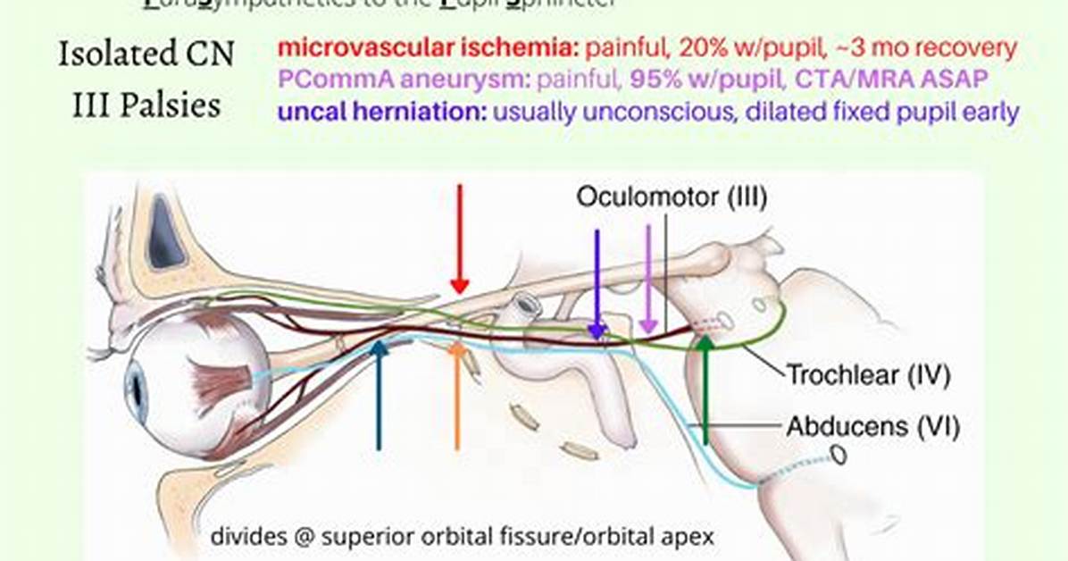
The oculomotor nerve exits the front part of the brainstem, between the midbrain and the pons. From here, cranial nerve 3 moves through the subarachnoid space, traveling to the eyes at the front of the skull. The subarachnoid space is an area between layers of brain matter. It allows cranial nerves, blood vessels and cerebrospinal fluid to travel through the brain.
The oculomotor nerve exits the brain by going through its outermost protective layer (dura mater). It enters the cavernous sinus, which is a hollow space behind the nose. As the nerve moves to the front part of the cavernous sinus, it splits into two branches: the superior branch and the inferior branch.
As they exit the cavernous sinus, both branches enter the orbit (eye socket) via the superior orbital fissure, which is a large hole behind each eye. Before reaching their destination, the two branches divide further to innervate (provide function to) the different muscles in and around the eye.
The superior branch splits to innervate the:
The inferior branch divides to innervate the:
Inferior oblique
Medial rectus
Inferior rectus
Short ciliary nerves
SEE RELATED: Optic nerve

